Department of Anatomy
Keeping abreast of improvements to the medical curriculum as envisaged in the CBME 2019 guidelines, Anatomy is now taught in a manner that makes it clinically relevant and facilitates integration with other disciplines. Subcomponents of the discipline include general anatomy, gross anatomy, embryology, histology, clinical anatomy, neuroanatomy, genetics, osteology, surface marking and radiological anatomy which are covered in a manner that not only provides sufficient scientific foundation but also makes them functionally relevant and of clinical significance.
The Department of Anatomy at PKDIMS teaches Anatomy to MBBS students in their first year, with teaching consisting of lecture classes, practical classes like dissection and histology lab practicals, small group discussion, self directed learning, early clinical exposure, vertical and horizontal integration and AETCOM. Phase one medical students receive the most comprehensive exposure to Human Anatomy over one year through lectures, small group discussions and supervised practical classes that provide hands on training through dissection of cadavers (for study of gross anatomy) as well as microscope slides for the study of histology. The study of clinical cases makes the study of Anatomy highly contextual. State of the art museum resources together with multimedia provide opportunities for references as well as self-directed learning.
We at department of Anatomy, PKDIMS plan to lay the foundations for a new approach to learning Anatomy by making this difficult subject engaging, stimulating and fun, and by helping students to be pro-active learners. Our role as Anatomy teachers is not to teach students anatomical facts, but rather to teach them the learning skills that will serve them throughout their student and professional lives. We hope to increase their cognitive learning skills and make them more self-directed in their learning. By experimenting with the new perspectives on traditional methods of teaching anatomy, we hope to provide a much more engaging, motivating, inspiring and enjoyable environment for student learning. When used in conjunction with traditional dissection, these innovative approaches appear to provide motivation that leads to deeper learning.
GOAL
The broad goal of teaching of undergraduate students in Anatomy aims at providing a comprehensive knowledge of the gross and microscopic structure and development of human body, which will serve as a basis for understanding the clinical correlation of organs or structures involved and the anatomical basis for the disease presentations.
OBJECTIVES
At the end of the course the students shall be able to:
Knowledge
- Comprehend the normal disposition, clinically relevant interrelationships, and functional and cross-sectional anatomy of the various structures in the body.
- Identify the microscopic structure and correlate elementary ultra-structure of various organs and tissues.
- Demonstrate knowledge of the basic principles and sequential development of the organs and systems; recognize the critical stages of development and the effects of common teratogens, genetic mutations, and environmental hazards. He /She shall be able to explain the developmental basis of the major variations and abnormalities.
Skills
At the end of the course the student shall be able to:
- Identify and locate all the structures of the body and mark the topography of the living anatomy.
- Identify the various organs and tissues under the microscope.
- State the principles of Karyotyping and identify the gross congenital anomalies.
- State principles of newer imaging techniques and interpretation of Ultrasonography, CT, MRI etc.
Integration
- By integrated teaching with other Phase I , Phase II and Phase III subjects students shall be able to comprehend the regulation and integration of the functions of the organs and systems in the body and thus interpret the anatomical basis of human body.
- Sensitize teachers about new concepts in teaching and assessment methods.
- Develop knowledge and clinical skills required for performing the role of competent and effective teacher, administrator, researcher and mentor.
- Update knowledge using modern information and research methodology tools.
OUR VISION
We help create a care plan that addresses your specific condition and we are here to answer all of your questions & acknowledge your concerns. Today the hospital is recognised as a world renowned institution, not only providing outstanding care and treatment, but improving the outcomes.
The Facilities
Museum
The excellent museum spread over 235 sq.mt., exhibits more than 235 dissected cadaveric and fetal specimens. The museum also displays sections of human body, radiographs, human skeletons, and developmental anatomy models. The museum collection is presented in several sections, e.g., Head and Neck, Thorax, Abdomen and Pelvis, Limbs, Sectional anatomy, Nervous system, and Embryology. New specimens and models are continuously added to the collection. Presently, genuine efforts are being made to promote the museum as a lively source of interaction for health science students and the public.
Dissection Hall
The 552 sq. mt. clean and brightly lit, hall ensures adequate space for more than 250 students at a time. The excellent cadaver preservation facilities include cold cabinets which can accommodate 16 cadavers. Formalin tanks for cadaver preservation can hold more than 40 cadavers and wash area is also available. Moreover, well preserved specimens of all parts of the human body are easily available to the students. The teaching-learning process is enhanced by normal X-ray, MRI, CT scan images and embryology models. The individual bones and articulated skeletons are also made accessible to the students during working hours. The dissection hall is well equipped with anthropometric instruments, X-ray view boxes and instruments essential for preparation of anatomical specimens, e.g., band saw, circular saw, brain knife etc.
Teaching Rooms
Two teaching rooms, each of 77.3 sq. mts., accommodating 75 students and equipped with appropriate audio-visual aids, add to the teaching ambience of the department. One of the teaching rooms is provided with state-of-the-art interactive LED panel, which facilitates a totally different new generation experience of visual and interactive Anatomy teaching.
Histology Lab
The department has a fully equipped histology unit spread over an area of 245 sq.mt. which can accommodate 90 students at a time. Students are provided with individual microscopes and slides during routine practical classes and a binocular microscope with CCTV attachment for the projection of slides for teaching. Coloured charts of labelled microanatomy sections of all tissues are displayed for students for reference during the routine practical sessions. The lab is also well equipped with tissue processing instruments, e.g., paraffin embedding bath, incubator, microtome etc. to make slides for teaching purposes. 90 state of the art microscopes and more than 5000 microscopic teaching slides are available in the histology lab to cater to the academic needs.
Embalming Room
Bodies received from appropriate sources after due processes are embalmed in this 17 sq. mts. room equipped with two embalming machines before being transferred to the storage tanks.
Storage Tanks
Three storage tanks of 3, 3 and 2 sq. mts. respectively are used for the storage of the embalmed bodies, namely, the cadavers. The number of cadavers will be proportionate to the number of students.
Lockers
150 lockers for storing student belongings during dissection hours are provided in the Anatomy Dissection Hall corridor.
Research laboratory
Research Lab is equipped with the instruments needed for the research activity in various fields of Anatomy like – Gross anatomy, embryology, histology, genetics, etc.
The available equipment include:
- Microtome Rotary Spencer Type
- Balance (Capacity – 6kg).
- Paraffin Embedding Bath.
- Hot Plate.
- Hot Air Oven.
- Refrigerators.
PhD in Anatomy under the guidance of Dr. P K Ramakrishnan
Two faculty members have successfully completed their PhD in Anatomy under the guidance of Dr. P K Ramakrishnan.
- Dr Akshara V R - Comparison of histomorphological changes in placenta with angiogenic markers in hypertensive disorders of pregnancy - A cross sectional study in Kerala.
- Dr Seema Valsalan - A comparative cross-sectional study on the morphological histological and immuno histochemical changes of umbilical cord in gestational Diabetes Mellitus and normal pregnancy in Kerala.
- Several ICMR short term studentship programs have been completed and are undergoing in the department.
Departmental Library
The departmental library consists of an excellent collection of books on Human Anatomy. In addition to these, standard reference books in gross anatomy, histology, embryology, neuroanatomy, osteology, surface marking, clinical anatomy, general anatomy, genetics and radiological anatomy are also available. This also facilitates the learning of clinical anatomy correlation with other clinical areas.
Pre Clinical Departments
Emergency Cases
Please feel welcome to contact our friendly reception staff with any general or medical enquiry call us.
Opening Hours
COVID-19 Latest Update
Our Medical HOD Professors
staffs all have exceptional experience and trained skills under various
medical departments and its treatment.
Richard Muldoone
Michael Brian
Maria Andaloro
Faculties
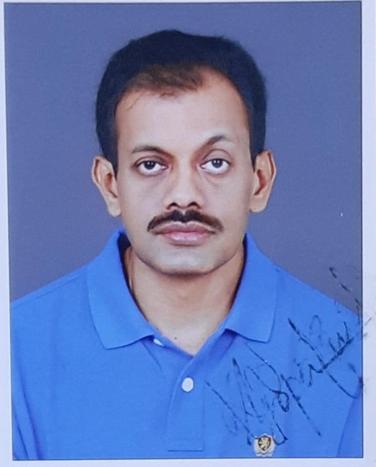
Dr. P. K. Ramakrishnan
MD
Professor
Reg.No: TCMC 24057
Dr. Benjamin. W
MD
Professor
Reg.No: TCMC 85068
Dr.Seema Valsalan E
MSc, PhD
Associate Professor
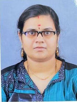
Dr. Aswathy V A
MD
Assistant Professor
Reg.No: TCMC 57562
Dr. Swaliha
MBBS
Tutor
Reg.No: TCMC 93183
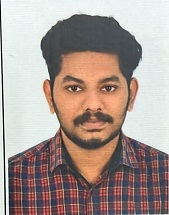
Dr. Abhil K Sathyan
MBBS
Tutor
Reg.No: TCMC 96298
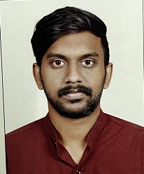
Dr. Adharv M R
MBBS
Tutor
Reg.No: TCMC 97128
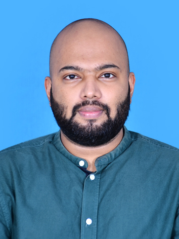
Dr. Siddique
MBBS
Tutor
Reg.No: TCMC 94713
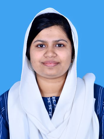
Dr. Faseetha
MBBS
Tutor
Reg.No: TCMC 91358
Dr. Ajmal Nizar
MBBS
Tutor
Reg.No: KSMC 90331
Dr. Nithin P.S
MBBS
Tutor
Reg.No: KSMC 96324
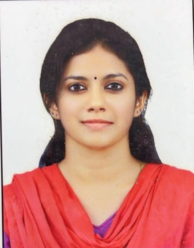
Dr. Anjali C Balagopalan
MBBS
Tutor
Reg.No: TCMC 70160
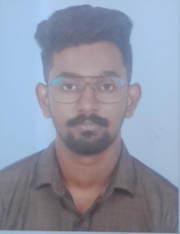
Dr. Anandhu V
MBBS
Tutor
Reg.No: KSMC 96159
Dr. Heaven Jose
MBBS, MD
Assistant Professor
Reg.No: TCMC 60926
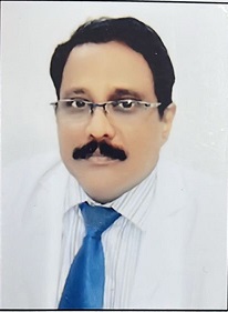
Dr. Nakka Vikas
MBBS, MD
Assistant Professor
Reg.No: APMC 17119
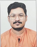
Dr. Karthik P
MBBS, MD
Tutor
Reg.No: KSMC 88794
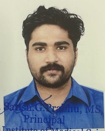
Dr. Karan S Kumar
MBBS
Tutor
Reg.No: KSMC 97392
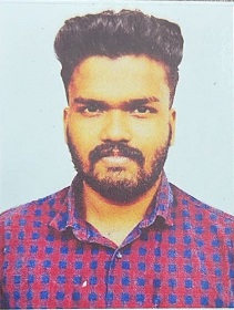
Dr. Sourav Bal P J
MBBS
Tutor
Reg.No: KSMC 96846
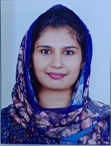
Dr. Baby Hind P
MBBS
Tutor
Reg.No: KSMC 92926
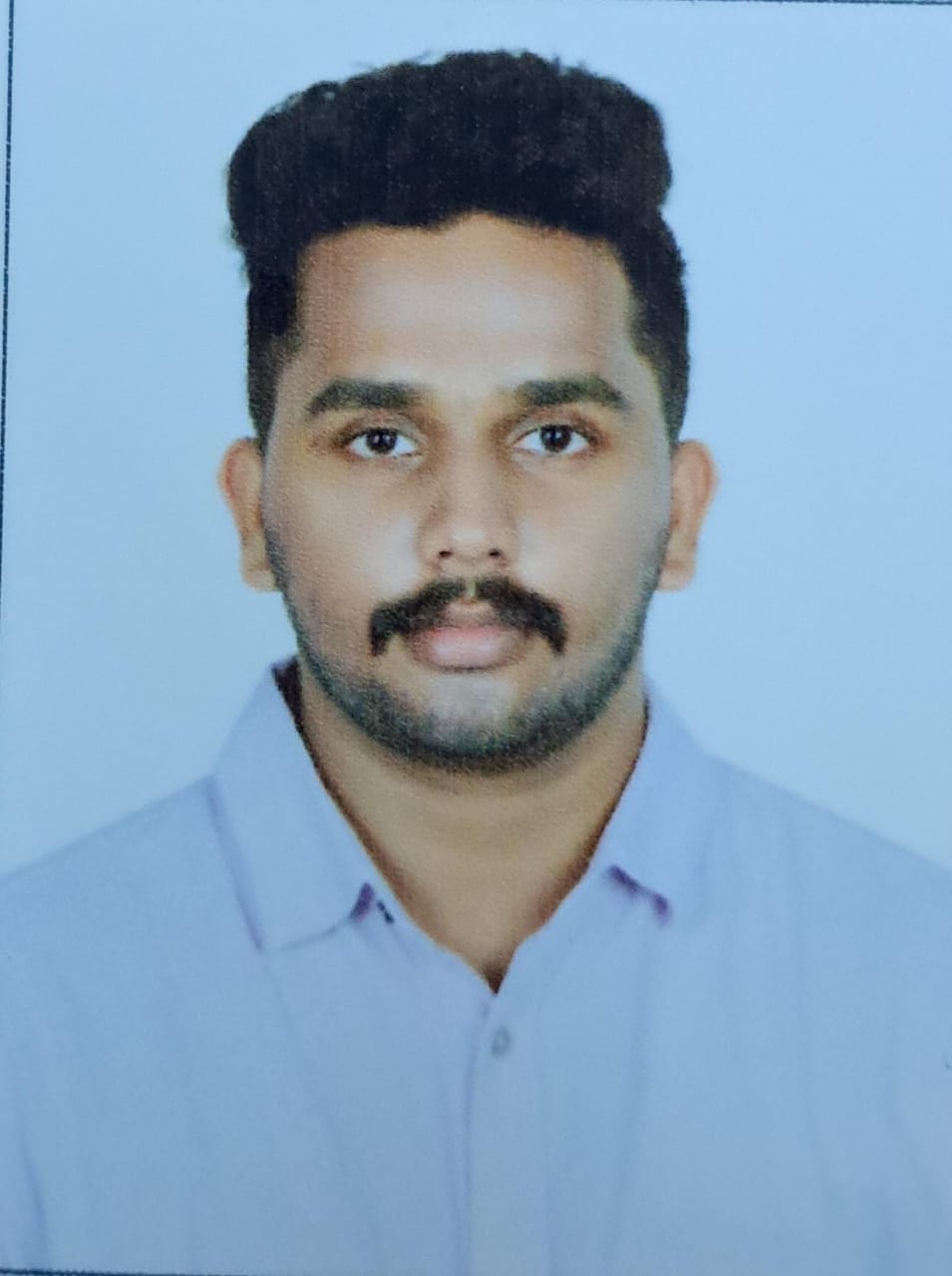
Dr. Arjun
MBBS
Tutor
Reg.No: KSMC 97514
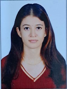
Dr. P M Sreelakhmi
MBBS
Tutor
Reg.No: KSMC 90237
Dr. Gayathri Sethurajan
MBBS
Tutor
Reg.No: 179437 - TMC
Dr. Someshwar C D
MBBS
Tutor
Reg.No: 165295 - TMC


